New images show clitoris anatomy in 3D
3D Imaging Unveils Clitoral Nerve System’s Complexity
A groundbreaking 3D study has shed light on the intricate nerve network of the clitoris, addressing longstanding gaps in medical understanding of female anatomy. For decades, the clitoris has been largely overlooked, with its size, location, and structure often misunderstood. This research, conducted by a team led by neuroscientist Ju Young Lee at Amsterdam University Medical Center, aims to correct that by revealing the full extent of the organ’s neural architecture.
Revealing the Clitoris’s Hidden Structure
Previously, the clitoris was commonly reduced to its visible tip, a small nub at the top of the vulva. However, the study shows that most of the organ lies internally, extending beneath the pubic bone and surrounding the vaginal opening. Its erectile tissue fills with blood during arousal, making it a key player in sexual response. Despite this, medical professionals have struggled to explain its anatomy with confidence, unlike the more widely studied male counterpart, the penis.
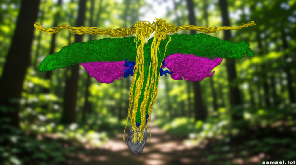
The research team utilized synchrotron radiation—a high-resolution X-ray imaging technique—to map the clitoris in microscopic detail. Unlike conventional MRI scans, which highlight broader anatomical features, this method allows for precise visualization of fine nerve pathways. The results demonstrate that the clitoris’s sensory nerves form a complex, tree-like branching pattern, rather than tapering as once believed.
“For the first time, the full trajectory of the terminal nerve branches of the clitoris has been mapped in three dimensions,” said Georga Longhurst, division lead of Anatomy and Physiology at the University of London. “Previous dissections and MRI studies showed these nerves before, but never to this level of detail.”
The findings also reveal that some nerve branches extend beyond the glans into the clitoral hood and even reach the mons pubis, the fatty tissue over the pubic bone. This comprehensive view of the clitoris’s nerve system challenges earlier assumptions and provides a clearer picture of its role in sexual sensation.
From Curiosity to Clinical Relevance
Lee, who initially focused on the brain, shifted her attention to peripheral nerve systems after noticing a lack of research on gynecological nerves. At a European neuroscience conference, she asked if anyone was examining how clitoral nerves communicate with the brain. A panelist’s surprise response—“Oh, I’d never thought about that”—prompted her to pursue the study further.
As part of the international Human Organ Atlas Hub, Lee’s work contributes to a project that maps the human body using advanced imaging. The clitoris, often considered a small external organ, is now shown to be a large and complex structure, comparable in size to the penis. When internal components are included, its total length measures up to 8 to 12 centimeters (3.1 to 4.7 inches), a detail that only gained traction in the late 1990s and early 2000s thanks to Australian urologist Helen O’Connell.
O’Connell’s MRI studies were pivotal in redefining the clitoris as a significant anatomical structure, not merely a tiny appendage. Lee’s latest research builds on this, offering surgeons a clearer understanding of the clitoral anatomy to minimize nerve damage during procedures. The preprint publication has already sparked interest in the medical community, underscoring the study’s practical implications for clinical practice.